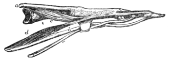
extensor communis between the heads of the metacarpal bones, while, rising from the shafts of these bones, are the three palmar and four dorsal interosseous muscles (fig. 9, e) which also are inserted into the extensor tendons. The two outer lumbricals and the thenar muscles are supplied by the median nerve; all the other hand muscles by the ulnar.
Muscles of the Lower Extremity.—On the front of the thigh the quadriceps extensor muscles are the most important: there are four of these, the rectus femoris (fig. 1) with its straight and reflected heads rising from just above the acetabulum, the crureus, deep to this, from the front of the femur, and the vastus externus and internus wrapping round the femur on each side from the linea aspera. All these are inserted into the patella, or rather the patella is a sesamoid bone developed where their common tendon passes round the lower end of the femur when the knee is bent.
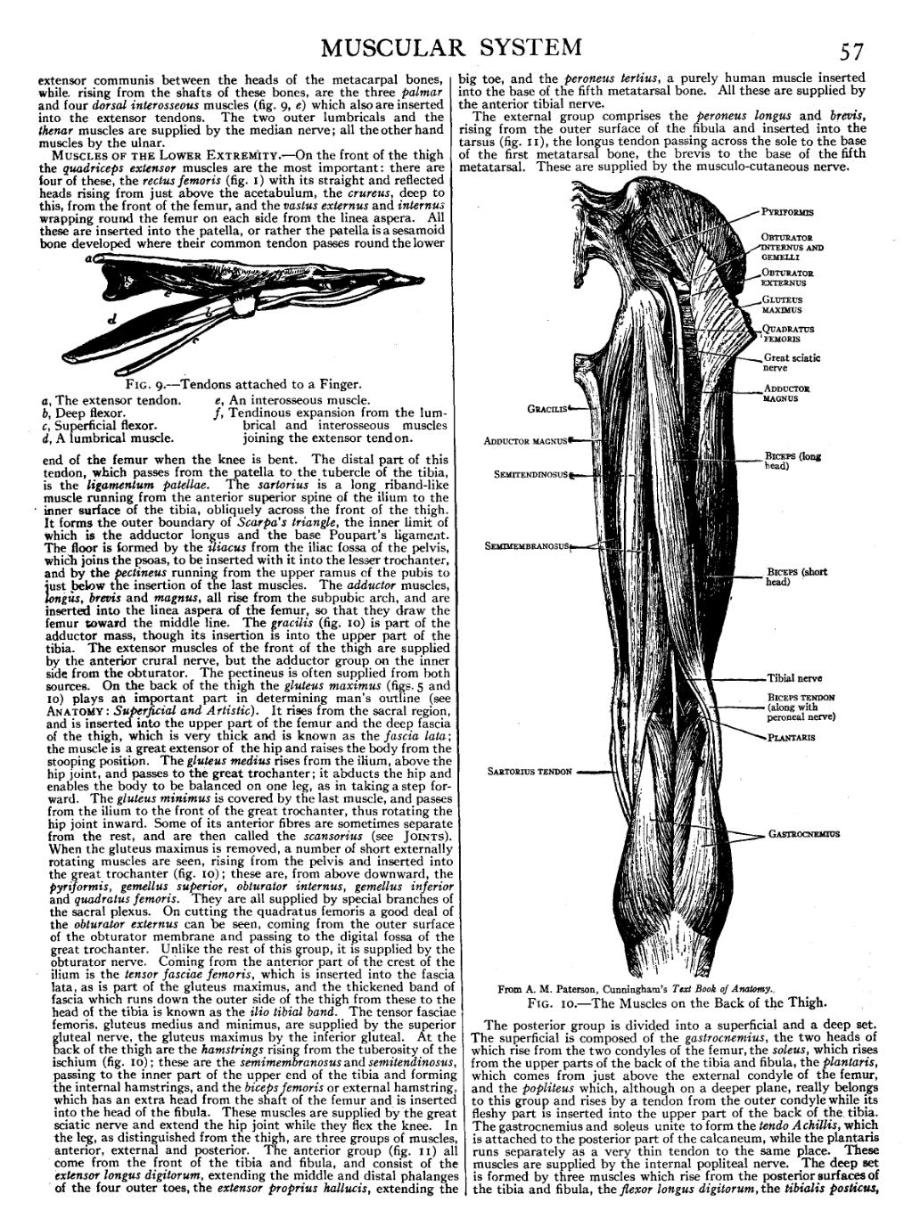
The distal part of this tendon, which passes from the patella to the tubercle of the tibia, is the ligamentum patellae. The sartorius is a long riband-like muscle running from the anterior superior spine of the ilium to the inner surface of the tibia, obliquely across the front of the thigh. It forms the outer boundary of Scarpa’s triangle, the inner limit of which is the adductor longus and the base Poupart's ligament. The floor is formed by the iliacus from the iliac fossa of the pelvis, which joins the psoas, to be inserted with it into the lesser trochanter, and by the pectineus running from the upper ramus of the pubis to just below the insertion of the last muscles. The adductor muscles, longus, brevis and magnus, all rise from the subpubic arch, and are inserted into the linea aspera of the femur, so that they draw the femur toward the middle line. The gracilis (fig. 10) is part of the adductor mass, though its insertion is into the upper part of the tibia. The extensor muscles of the front of the thigh are supplied by the anterior crural nerve, but the adductor group on the inner side from the obturator. The pectineus is often supplied from both sources. On the back of the thigh the gluteus maximus (figs. 5 and 10) plays an important part in determining man’s outline (see Anatomy: Superficial and Artistic). It rises from the sacral region, and is inserted into the upper part of the femur and the deep fascia of the thigh, which is very thick and is known as the fascia lata; the muscle is a great extensor of the hip and raises the body from the stooping position. The gluteus medius rises from the ilium, above the hip joint, and passes to the great trochanter; it abducts the hip and enables the body to be balanced on one leg, as in taking a step forward. The gluteus minimus is covered by the last muscle, and passes from the ilium to the front of the great trochanter, thus rotating the hip joint inward. Some of its anterior fibres are sometimes separate from the rest, and are then called the scansorius (see Joints). When the gluteus maximus is removed, a number of short externally rotating muscles are seen, rising from the pelvis and inserted into the great trochanter (fig. 10); these are, from above downward, the pyriformis, gemellus superior, obturator internus, gemellus inferior and quadratus femoris. They are all supplied by special branches of the sacral plexus. On cutting the quadratus femoris a good deal of the obturator externus can be seen, coming from the outer surface of the obturator membrane and passing to the digital fossa of the great trochanter. Unlike the rest of this group, it is supplied by the obturator nerve. Coming from the anterior part of the crest of the ilium is the tensor fasciae femoris, which is inserted into the fascia lata, as is part of the gluteus maximus, and the thickened band of fascia which runs down the outer side of the thigh from these to the head of the tibia is known as the ilio tibial band. The tensor fasciae femoris. gluteus medius and minimus, are supplied by the superior gluteal nerve, the gluteus maximus by the inferior gluteal. At the back of the thigh are the hamstrings rising from the tuberosity of the ischium (fig. 10); these are the semimembranosus and semitendinosus, passing to the inner part of the upper end of the tibia and forming the internal hamstrings, and the biceps femoris or external hamstring, which has an extra head from the shaft of the femur and is inserted into the head of the fibula. These muscles are supplied by the great sciatic nerve and extend the hip joint while they flex the knee. In the leg, as distinguished from the thigh, are three groups of muscles, anterior, external and posterior. The anterior group (fig. 11) all come from the front of the tibia and fibula, and consist of the extensor longus digitorum, extending the middle and distal phalanges of the four outer toes, the extensor proprius hallucis, extending the big toe, and the peroneus tertius, a purely human muscle inserted into the base of the fifth metatarsal bone. All these are supplied by the anterior tibial nerve.
The external group comprises the peroneus longus and brevis, rising from the outer surface of the fibula and inserted into the tarsus (fig. 11), the longus tendon passing across the sole to the base of the first metatarsal bone, the brevis to the base of the fifth metatarsal. These are supplied by the musculo-cutaneous nerve.
 |
| From A. M. Paterson, Cunningham's Text Book of Anatomy. |
Fig. 10.—The Muscles on the Back of the Thigh. |

