Jump to content

Report on the Radiolaria/Plates6

From Wikisource
1584887Report on the Radiolaria — Plates 51-60Ernst Haeckel

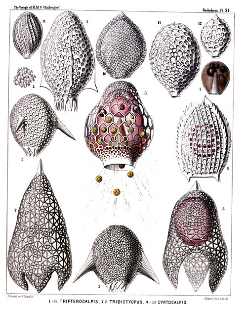
PLATE 51.

Legion NASSELLARIA.

Order CYRTOIDEA.

Families Tripocalpida, Phænocalpida et Cyrtocalpida.


PLATE 51.
Tripocalpida, Phænocalpida et Cyrtocalpida.
Diam. Page.
Fig. 1. Tripterocalpis phylloptera, n. sp., × 400 1138
Fig. 2. Tripterocalpis conoptera, n. sp., × 300 1138
Fig. 3. Tripterocalpis ogmoptera, n. sp., × 300 1138
Fig. 4. Tripterocalpis ogmoptera, n. sp., × 500 1138
A group of confluent pores, more enlarged.
Fig. 5. Tripterocalpis ogmoptera, n. sp., × 300 1138
Central capsule. In the centre the striate podoconus, above it four oil-globules, to the right the nucleus.
Fig. 6. Tripocalpis triserrata, n. sp., × 600 1136
Fig. 7. Tridictyopus conicus, n. sp., × 300 1145
Fig. 8. Tridictyopus vatillum, n. sp., × 400 1145
Fig. 9. Cyrtophormis spiralis, n. sp., × 400 1166
Fig. 10. Archicorys ovata, n. sp., × 300 1185
Fig. 11. Cyrtocalpis gromia, n. sp., × 400 1188
Fig. 12. Archicorys microstoma, n. sp., × 400 1185
Fig. 13. Cyrtocalpis urceolus, n. sp., × 500 1186
The ovate central capsule exhibits in the lower half the podoconus, in the upper half the spherical nucleus and three oil-globules. Between the capsule and the shell numerous xanthellæ, partly protruded through the shell-mouth along the radiating pseudopodia.
Plate 51.
Plate 51.


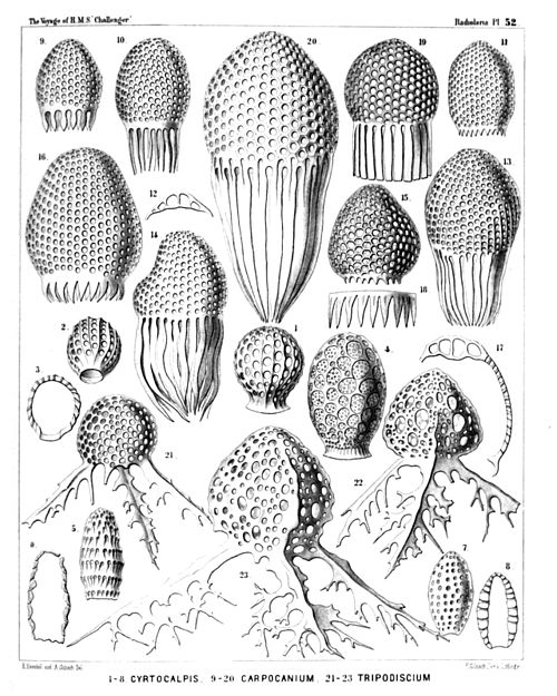
PLATE 52.

Legion NASSELLARIA.

Order CYRTOIDEA.

Families Tripocalpida, Phænocalpida, Cyrtocalpida et Anthocyrtida.


PLATE 52.
Tripocalpida, Phænocalpida, Cyrtocalpida et Anthocyrtida.
Diam. Page.
Fig. 1. Cyrtophormis pila, n. sp., × 300 1165
Fig. 2. Cyrtophormis ærostatica, n. sp., × 300 1166
Fig. 3. Cyrtophormis ærostatica, n. sp., × 300 1166
Longitudinal section.
Fig. 4. Cyrtocalpis sethopora, n. sp., × 600 1187
Fig. 5. Cyrtocalpis lithomitra, n. sp., × 400 1187
Fig. 6. Cyrtocalpis lithomitra, n. sp., × 400 1187
Longitudinal section.
Fig. 7. Cyrtocalpis compacta, n. sp., × 400 1187
Fig. 8. Cyrtocalpis compacta, n. sp., × 400 1187
Longitudinal section.
Fig. 9. Carpocanistrum flosculum, n. sp., × 400 1171
Fig. 10. Carpocanistrum cephalum, n. sp., × 300 1171
Fig. 11. Carpocanistrum evacuatum, n. sp., × 400 1172
Fig. 12. Carpocanium verecundum, n. sp., × 400 1284
Vertical section through the top of the shell.
Fig. 13. Carpocanium verecundum, n. sp., × 400 1284
Fig. 14. Carpocanium irregulare, n. sp., × 400 1284
Fig. 15. Carpocanium hexagonale, n. sp., × 400 1282
Fig. 16. Carpocanium peristomium, n. sp., × 500 1283
Fig. 17. Carpocanium peristomium, n. sp., × 500 1283
Vertical section.
Fig. 18. Carpocanium trepanium, n. sp., × 600 1282
Peristome.
Fig. 19. Carpocanium petalospyris, n. sp., × 300 1283
Fig. 20. Carpocanium virgineum, n. sp., × 600 1285
Fig. 21. Tripodiscium sphærocephalum, n. sp., × 400 1144
Fig. 22. Tripodiscium tristylospyris, n. sp. (vel Tristylospyris tripodiscium), × 600 1143
Fig. 23. Tripodiscium ramosum, n. sp. (vel Tristylospyris ramosa), × 600 1144
Plate 52.
Plate 52.


PLATE 53.

Legion NASSELLARIA.

Orders SPYROIDEA et CYRTOIDEA.

Families Zygospyrida, Tripocalpida, Phænocalpida et Cyrtocalpida.


PLATE 53.
Zygospyrida, Tripocalpida, Phænocalpida et Cyrtocalpida.
Diam. Page.
Fig. 1. Archicapsa triforis, n. sp., × 300 1191
Lateral view.
Fig. 2. Archicapsa triforis, n. sp., × 300 1191
Basal view.
Fig. 3. Halicapsa triglochin, n. sp., × 200 1190
Lateral view.
Fig. 4. Halicapsa triglochin, n. sp., × 200 1191
Basal view.
Fig. 5. Halicapsa hystrix, n. sp., × 200 1191
Lateral view.
Fig. 6. Halicapsa hystrix, n. sp., × 200 1191
Basal view.
Fig. 7. Cantharospyris platybursa, n. sp. (vel Platybursa compressa), × 400 1051
Fig. 8. Tessarospyris clathrobursa, n. sp. (vel Clathrobursa dictyopus), × 400 1045
Fig. 9. Peridium spinipes, n. sp., × 500 1154
Fig. 10. Peridium palmipes, n. sp., × 500 1154
Fig. 11. Archiscenium quadrispinum, n. sp., × 500 1150
In the spherical central capsule the dark nucleus is visible.
Fig. 12. Euscenium eucolpium, n. sp., × 500 1147
Fig. 13. Cladoscenium ancoratum, n. sp., × 400 1149
Fig. 14. Pteroscenium pinnatum, n. sp., × 400 1152
Lateral view.
Fig. 15. Pteroscenium pinnatum, n. sp., × 400 1152
Vertical section.
Fig. 16. Pteroscenium pinnatum, n. sp., × 400 1152
Basal view.
Fig. 17. Calpophæna hexarrhabda, n. sp., × 400 1176
Fig. 18. Calpophæna hexarrhabda, n. sp., × 400 1176
Basal plate.
Fig. 19. Tetraspyris tetracorethra, n. sp., × 400 1044
With the four-lobed central capsule, in each lobe an oil-globule.
Fig. 20. Tetraspyris tetracorethra, n. sp., × 800 1044
Shell more enlarged.
Plate 53.
Plate 53.


PLATE 54.

Legion NASSELLARIA.

Order CYRTOIDEA.

Families Phænocalpida, Cyrtocalpida, Anthocyrtida et Sethocyrtida.


PLATE 54.
Phænocalpida, Cyrtocalpida, Anthocyrtida et Sethocyrtida.
Diam. Page.
Fig. 1. Bathropyramis quadrata, n. sp., × 300 1159
Fig. 2. Sethopyramis quadrata, n. sp., × 300 1254
Fig. 3. Bathropyramis trapezoides, n. sp., × 300 1160
Fig. 4. Bathropyramis ramosa, n. sp., × 300 1161
Fig. 5. Peripyramis circumtexta, n. sp., × 300 1162
Fig. 6. Plectopyramis dodecomma, n. sp., × 400 1258
Fig. 7. Cinclopyramis infundibulum, n. sp., × 300 1161
Fig. 8. Plectopyramis trapezomma, n. sp., × 400 1258
Fig. 9. Cornutella hexagona, n. sp., × 400 1180
Fig. 10. Cornutella sethoconus, n. sp., × 400 1180
Fig. 11. Sethoconus orthoceras, n. sp., × 400 1294
Fig. 12. Sethoconus bimarginatus, n. sp., × 400 1295
Plate 54.
Plate 54.


PLATE 55.

Legion NASSELLARIA.

Order CYRTOIDEA.

Families Phænocalpida, Anthocyrtida et Sethocyrtida.


PLATE 55.
Phænocalpida, Anthocyrtida et Sethocyrtida.
Diam. Page.
Fig. 1. Sethoconus facetus, n. sp. (vel Phlebarachnium facetum), × 300 1296
Upper part of the shell.
Fig. 2. Sethoconus venosus, n. sp. (vel Phlebarachnium venosum), × 250 1297
Shell including the four-lobed central capsule.
Fig. 3. Sethophormis aurelia, n. sp. (vel Leptarachnium aurelia), × 100 1248
Shell seen from above.
Fig. 4. Sethophormis aurelia, n. sp., × 400 1248
Cephalis more enlarged, with the enclosed four-lobed central capsule.
Fig. 5. Cladarachnium ramosum, n. sp., × 300 1165
Apical view.
Fig. 6. Cladarachnium ramosum, n. sp., × 70 1165
Lateral view.
Fig. 7. Bathropyramis interrupta, n. sp., × 300 1160
Apical part of the shell, from above.
Fig. 8. Litharachnium araneosum, n. sp., × 300 1163
Apical part of the shell, from above.
Fig. 9. Litharachnium epeira, n. sp., × 500 1164
Oblique view of the shell.
Fig. 10. Litharachnium araneosum, n. sp., × 50 1163
Lateral view.
Fig. 11. Periarachnium periplectum, n. sp., × 500 1297
Shell enclosing the trilobed central capsule.
Plate 55.
Plate 55.


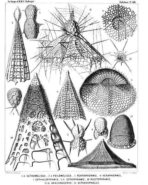
PLATE 56.

Legion NASSELLARIA.

Order CYRTOIDEA.

Families Tripocyrtida, Anthocyrtida et Sethocyrtida.


PLATE 56.
Tripocyrtida, Anthocyrtida et Sethocyrtida.
Diam. Page.
Fig. 1. Lithomelissa bütschlii, n. sp. (vel Sethomelissa bütschlii), × 400 1207
Fig. 2. Lithomelissa decacantha, n. sp. (vel Sethomelissa decacantha), × 400 1208
Fig. 3. Psilomelissa calvata, n. sp., × 400 1209
The cephalis alone, with the three collar beams.
Fig. 4. Lychnodictyum scaphopodium, n. sp., × 400 1231
Fig. 5. Sethophormis pentalactis, n. sp. (vel Pentaphormis pentalactis), × 400 1244
Oblique view of the shell, from below.
Fig. 6. Sethophormis hexalactis, n. sp. (vel Hexaphormis hexalactis), × 400 1245
Central part of the shell, with the cortinar septum.
Fig. 7. Sethopyramis enneactis, n. sp. (vel Cephalopyramis enneactis), × 400 1254
Fig. 8. Plectopyramis polypleura, n. sp. (vel Sethopyramis polypleura), × 200 1260
Fig. 9. Sethophormis eupilium, n. sp. (vel Craspedilium eupilium), × 400 1247
Fig. 10. Plectopyramis spongiosa, n. sp. (vel Spongopyramis spongiosa), × 400 1261
Fig. 11. Arachnocorys araneosa, n. sp., × 500 1266
Fig. 12. Sethophormis dodecaster, n. sp. (vel Astrophormis dodecaster), × 200 1248
Fig. 13. Sethocephalus eucecryphalus, n. sp., × 400 1298
Plate 56.
Plate 56.


PLATE 57.

Legion NASSELLARIA.

Order CYRTOIDEA.

Families TRIPOCYRTIDA, ANTHOCYRTIDA et SETHOCYRTIDA.


PLATE 57.
TRIPOCYRTIDA, ANTHOCYRTIDA et SETHOCYRTIDA.
Diam. Page.
Fig. 1. Dicolocapsa microcephala, n. sp., × 400 1312
Fig. 2. Sethocapsa pyriformis, n. sp., × 300 1310
Fig. 3. Lithopera ananassa, n. sp., × 500 1234
Fig. 4. Sethamphora favosa, n. sp. (vel Cryptoprora favosa), × 400 1252
Fig. 5. Sethamphora microstoma, n. sp. (vel Cryptoprora microstoma), × 300 1252
Fig. 6. Clistophæna hexolena, n. sp., × 300 1287
Fig. 7. Clistophæna armata, n. sp., × 300 1288
Fig. 8. Clathromitra pterophormis, n. sp., × 400 1219
Fig. 9. Sethophormis rotula, n. sp. (vel Enneaphormis rotula), × 400 1246
Fig. 10. Dictyophimus sphærocephalus, n. sp., × 400 1195
Fig. 11. Peromelissa phalacra, n. sp., × 400 1236
Fig. 12. Peromelissa calva, n. sp., × 300 1237
Fig. 13. Sethochytris triconiscus, n. sp., × 300 1239
Fig. 14. Micromelissa bombus, n. sp., × 300 1235
Plate 57.
Plate 57.


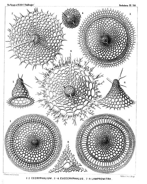
PLATE 58.

Legion NASSELLARIA.

Order CYRTOIDEA.

Families Tripocyrtida, Sethocyrtida, Phormocyrtida et Theocyrtida.


PLATE 58.
Tripocyrtida, Sethocyrtida, Phormocyrtida et Theocyrtida.
Diam. Page.
Fig. 1. Cecryphalium sestrodiscus, n. sp., × 400 1399
Apical view.
Fig. 2. Cecryphalium lamprodiscus, n. sp., × 400 1398
Apical view.
Fig. 3. Clathrocyclas coscinodiscus, n. sp., × 400 1389
Apical view.
Fig. 4. Clathrocyclas coscinodiscus, n. sp., × 700 1389
The cephalis alone, with the two horns.
Fig. 5. Clathrocyclas semeles, n. sp., × 400 1388
Lateral view.
Fig. 6. Sethoconus capreolus, n. sp., × 400 1291
Lateral view.
Fig. 7. Lampromitra quadricuspis, n. sp., × 400 1214
Apical view.
Fig. 8. Lampromitra furcata, n. sp., × 400 1215
The collar septum after removal of the cephalis.
Fig. 9. Lampromitra dendrocorona, n. sp., × 400 1216
Apical view.
Plate 58.
Plate 58.


PLATE 59.

Legion NASSELLARIA.

Order CYRTOIDEA.

Families Tripocyrtida, Podocyrtida et Phormocyrtida.


PLATE 59.
Tripocyrtida, Podocyrtida et Phormocyrtida.
Diam. Page.
Fig. 1. Lampromitra huxleyi, n. sp., × 400 1215
Fig. 2. Amphiplecta callistoma, n. sp., × 400 1224
Fig. 3. Corocalyptra agnesæ, n. sp., × 400 1323
Fig. 4. Corocalyptra emmæ, n. sp., × 400 1323
The shell encloses the trilobate central capsule, with the trilobate nucleus.
Fig. 5. Clathrocyclas cassiopejæ, n. sp., × 400 1390
Fig. 6. Clathrocyclas alcmenæ, n. sp., × 400 1388
Fig. 7. Clathrocyclas latonæ, n. sp., × 400 1389
Apical view.
Fig. 8. Diplocylas bicorona, n. sp., × 400 1392
Fig. 9. Clathrocyclas ionis, n. sp., × 400 1389
Fig. 10. Corocalyptra elisabethæ, n. sp., × 400 1323
Oblique apical view of the shell, with the quadrilobate central capsule enclosed.
Fig. 11. Clathrocyclas europæ, n. sp., × 400 1388
Apical view of the shell, after removal of the cephalis.
Fig. 12. Clathrocyclas europæ, n. sp., × 400 1388
Central capsule, seen from above, with the quadrilobate nucleus.
Fig. 13. Clathrocyclas danaës, n. sp., × 300 1388
Vertical section through the cephalis and the quadrilobate central capsule, with the quadrilobate nucleus.
Fig. 14. Clathrocyclas danaës, n. sp., × 300 1388
Apical view of the shell.
Plate 59.
Plate 59.


PLATE 60.

Legion NASSELLARIA.

Order CYRTOIDEA.

Family Tripocyrtida.


PLATE 60.
Tripocyrtida.
Diam. Page.
Fig. 1. Dictyophimus cienkowskii, n. sp. (vel Lamprotripus squarrosus), × 300 1200
Shell seen from the side.
Fig. 2. Dictyophimus bütschlii, n. sp. (vel Lamprotripus horridus), × 300 1201
Fig. 3. Dictyophimus hertwigii, n. sp. (vel Lamprotripus spinosus), × 400 1201
The cephalis of the shell includes the central capsule, with three lobes depending in the pyramidal thorax.
Fig. 4. Dictyophimus platycephalus, n. sp., × 400 1198
Central capsule with four thoracic lobes, each of which contains an oil-globule; kidney-shaped nucleus in the cephalic lobe.
Fig. 5. Dictyophimus platycephalus, n. sp., × 400 1198
Shell seen from the side.
Fig. 6. Dictyophimus brandtii, n. sp., × 300 1198
Shell seen from the base, with the four large pores of the collar septum, two minor jugular and two major cardinal pores.
Fig. 7. Lampromitra coronata, n. sp., × 400 1214
Shell seen from below, with the quadrilobate central capsule.
Fig. 7a. A portion of the shell-margin, × 800 1214
Fig. 8. Lampromitra arborescens, n. sp., × 400 1216
Shell from above.
Fig. 8a. The collar septum with the four crossed rods of the cortina, × 400 1216
Fig. 9. Tripocyrtis plectaniscus, n. sp., × 400 1202
Fig. 10. Tripocyrtis plagoniscus, n. sp., × 400 1201
Plate 60.
Plate 60.