(the eye-stalk and sessile lateral eyes of Arthropoda generally, exclusive of Peripatus).
(10) The forms assumed by special modification of the elements of the parapodium in the maxillae, labium, &c., of Hexapods, Chilopods, Diplopods, and of various Crustacea, deserve special enumeration, but cannot be dealt with without ample space and illustration.
It may be pointed out that the most radical difference presented in this list is that between appendages consisting of the corm alone without rami (Onychophora) and those with more or less developed rami (the rest of the Arthropoda). In the latter class we should distinguish three phases: (a) those with numerous and comparatively undeveloped rami; (b) those with three, or two highly developed rami, or with only one—the corm being reduced to the dimensions of a mere basal segment; (c) those reduced to a secondary simplicity (degeneration) by overwhelming development of one segment (e.g. the isolated gnathobase often seen as “mandible” and the genital operculum).
There is no reason to suppose that any of the forms of limb observed in Arthropoda may not have been independently developed in two or more separate diverging lines of descent.
Branchiae.—In connexion with the discussion of the limbs of Arthropods, a few words should be devoted to the gill-processes. It seems probable that there are branchial plumes or filaments in some Arthropoda (some Crustacea) which can be identified with the distinct branchial organs of Chaetopoda, which lie dorsal of the parapodia and are not part of the parapodium. On the other hand, we cannot refuse to admit that any of the processes of an Arthropod parapodium may become modified as branchial organs, and that, as a rule, branchial out-growths are easily developed, de novo, in all the higher groups of animals. Therefore, it seems to be, with our present knowledge, a hopeless task to analyse the branchial organs of Arthropoda and to identify them genetically in groups.
A brief notice must suffice of the structure and history of the Eyes, the Tracheae and the so-called Malpighian tubes of Arthropoda, though special importance attaches to each in regard to the determination of the affinities of the various animals included in this great sub-phylum.
The Eyes.—The Arthropod eye appears to be an organ of special character developed in the common ancestor of the Euarthropoda, and distinct from the Chaetopod eye, which is found only in the Onychophora where the true Arthropod eye is absent. The essential difference between these two kinds of eye appears to be that the Chaetopod eye (in its higher developments) is a vesicle enclosing the lens, whereas the Arthropod eye is a pit or series of pits into which the heavy chitinous cuticle dips and enlarges knobwise as a lens. Two distinct forms of the Arthropod eye are observed—the monomeniscous (simple) and the polymeniscous (compound). The nerve-end-cells, which lie below the lens, are part of the general epidermis. They show in the monomeniscous eye (see article Arachnida, fig. 26) a tendency to group themselves into “retinulae,” consisting of five to twelve cells united by vertical deposits of chitin (rhabdoms). In the case of the polymeniscous eye (fig. 23, article Arachnida) a single retinula or group of nerve-end-cells is grouped beneath each associated lens. A further complication occurs in each of these two classes of eye. The monomeniscous eye is rarely provided with a single layer of cells beneath its lens; when it is so, it is called monostichous (simple lateral eye of Scorpion, fig. 22, article Arachnida). More usually, by an infolding of the layer of cells in development, we get three layers under the lens; the front layer is the corneagen layer, and is separated by a membrane from the other two which, more or less, fuse and contain the nerve-end-cells (retinal layer). These eyes are called diplostichous, and occur in Arachnida and Hexapoda (fig. 24, article Arachnida).
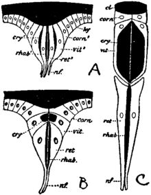
On the other hand, the polymeniscous eye undergoes special elaboration on its lines. The retinulae become elongated as deep and very narrow pits (fig. 12 and explanation), and develop additional cells near the mouth of the narrow pit. Those nearest to the lens are the corneagen cells of this more elaborated eye, and those between the original retinula cells and the corneagen cells become firm and transparent. They are the crystalline cells or vitrella (see Watase, 7). Each such complex of cells underlying the lenticle of a compound eye is called an “ommatidium”; the entire mass of cells underlying a monomeniscous eye is an “ommataeum.” The ommataeum, as already stated, tends to segregate into retinulae which correspond potentially each to an ommatidium of the compound eye. The ommatidium is from the first segregate and consists of few cells. The compound eye of the king-crab (Limulus) is the only recognized instance of ommatidia in their simplest state. Each can be readily compared with the single-layered lateral eye of the scorpion. In Crustacea and Hexapoda of all grades we find compound eyes with the more complicated ommatidia described above. We do not find them in any Arachnida.
It is difficult in the absence of more detailed knowledge as to the eyes of Chilopoda and Diplopoda to give full value to these facts in tracing the affinities of the various classes of Arthropods. But they seem to point to a community of origin of Hexapods and Crustacea in regard to the complicated ommatidia of the compound eye, and to a certain isolation of the Arachnida, which are, however, traceable, so far as the eyes are concerned, to a distant common origin with Crustacea and Hexapoda through the very simple compound eyes (monostichous, polymeniscous) of Limulus.

|
Fig. 12.—Diagram to show the derivation of the unit or “ommatidium” of the compound eye of Crustacea and Hexapoda, C, from a simple monomeniscous monostichous eye resembling the lateral eye of a scorpion, A, or the unit of the compound lateral eye of Limulus (see article Arachnida, figs. 22 and 23). B represents an intermediate hypothetical form in which the cells beneath the lens are beginning to be superimposed as corneagen, vitrella and retinula, instead of standing side by side in horizontal series. The black represents the cuticular product of the epidermal cells of the ocular area, taking the form either of lens, cl, of crystalline body, cry, or of rhabdom, rhab; hy, hypodermis or epidermal cells; corn1, laterally-placed cells in the simpler stage, A, which like the nerve-end cells, vit1 and ret1, are corneagens or lens-producing; corn, specialized corneagen or lens-producing cells; vit1, potential vitrella cells with cry1, potential crystalline body now indistinguishable from retinula cells and rhabdomeres; vit, vitrella cell with cry, its contained cuticular product, the crystalline cone or body; ret1, rhab1, retinula cells and rhabdom of scorpion undifferentiated from adjacent cells, vit1; ret, retinula cell; rhab, rhabdom; nf, optic nerve-fibres. (Modified from Watase.) |
The Tracheae.—In regard to tracheae the very natural tendency of zoologists has been until lately to consider them as having once developed and once only, and therefore to hold that a group “Tracheata” should be recognized, including all tracheate Arthropods. We are driven by the conclusions arrived at as to the derivation of the Arachnida from branchiate ancestors, independently of the other tracheate Arthropods, to formulate the conclusion that tracheae have been independently developed in the Arachnidan class. We are also, by the isolation of Peripatus and the impossibility of tracing to it all other tracheate Arthropoda, or of regarding it as a degenerate offset from some one of the tracheate classes, forced to the conclusion that the tracheae of the Onychophora have been independently acquired. Having accepted these two conclusions, we formulate the generalization that tracheae can be independently acquired by various branches of Arthropod descent in adaptation to a terrestrial as opposed to an aquatic mode of life. A great point of interest therefore exists in the knowledge of the structure and embryology of tracheae in the different groups. It must be confessed that we have not such full knowledge on this head as could be wished for. Tracheae are essentially tubes like blood-vessels—apparently formed from the same tissue elements as blood-vessels—which contain air in place of blood, and usually communicate by definite orifices, the tracheal stigmata, with the atmosphere. They are lined internally by a cuticular deposit of chitin. In Peripatus and the Diplopods they consist of bunches of fine tubes which do not branch but diverge from one another; the chitinous lining is smooth. In the Hexapods and Chilopods, and the Arachnids (usually), they form tree-like branching structures, and their finest branches are finer than any blood-capillary, actually in some cases penetrating a single cell and supplying it with gaseous oxygen. In these forms the chitinous lining of the tubes is thickened by a close-set spiral ridge similar to the spiral thickening of the cellulose wall of the spiral vessels of plants. It is a noteworthy fact that other tubes in these same terrestrial Arthropoda—namely, the ducts of glands—are similarly strengthened by a chitinous cuticle, and that a spiral or annular thickening of the cuticle is developed in them also. Chitin is not exclusively an ectodermal product, but occurs also in cartilaginous skeletal plates of mesoblastic origin (connective tissue). The immediate cavities or pits into which the tracheal stigmata open appear to be in many cases ectodermic in sinkings, but there seems to be no reason (based on embryological observation) for regarding the tracheae as an ingrowth of the ectoderm. They appear, in fact, to be an air-holding modification of the vasifactive connective tissue. Tracheae are abundant just in proportion as blood-vessels become suppressed. They are reciprocally exclusive. It seems not improbable that they are two modifications of the same tissue-elements. In Peripatus the stigmatic pits at which the tracheae communicate with the atmosphere are scattered and not definite in their position. In other cases the stigmata are definitely paired and placed in a few segments or in several. It seems that we have to suppose that the vasifactive tissue of Arthropoda can readily take the form of air-holding instead of blood-holding tubes, and that this somewhat startling change in its character has taken place independently in several instances—viz. in the Onychophora, in more than one group of Arachnida, in Diplopoda, and again in the Hexapoda and Chilopoda.
The Malpighian Tubes.—This name is applied to the numerous fine caecal tubes of noticeable length developed from the proctodaeal