Jump to content

Report on the Radiolaria/Plates9

From Wikisource
1584908Report on the Radiolaria — Plates 81-90Ernst Haeckel

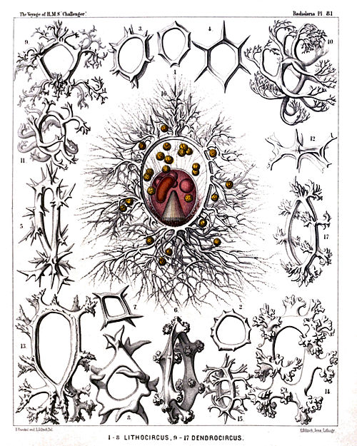
PLATE 81.

Legion NASSELLARIA.

Order STEPHOIDEA.

Family Stephanida.


PLATE 81.
Stephanida.
Diam. Page.
Fig. 1. Archicircus primordialis, n. sp., × 200 942
Fig. 2. Zygocircus polygonus, n. sp., × 200 947
Fig. 3. Zygocircus triquetrus, n. sp., × 300 947
Fig. 4. Archicircus hexacanthus, n. sp., × 300 942
Fig. 5. Zygocircus acacia, n. sp., × 300 947
Fig. 6. Lithocircus crambessa, n. sp., × 400 944
Fig. 7. Archicircus rhombus, n. sp., × 300 942
Fig. 8. Zygocircus pentagonus, n. sp., × 300 946
Fig. 9. Lithocircus quadricornis, n. sp., × 300 944
Fig. 10. Dendrocircus arborescens, n. sp., × 300 949
Fig. 11. Dendrocircus dodecancistra, n. sp., × 300 949
Fig. 12. Archicircus sexangularis, n. sp., × 300 943
Fig. 13. Dendrocircus elegans, n. sp., × 400 949
Fig. 14. Dendrocircus stalactites, n. sp., × 400 950
Fig. 15. Lithocircus decimalis, n. sp., × 300 944
Fig. 16. Lithocircus magnificus, n. sp., × 400 945
The ovate, red-coloured central capsule exhibits in the lower half the striate podoconus, in the upper half four oil-globules, and at the left the kidney-shaped nucleus. Numerous "yellow cells" or xanthellæ are scattered in the calymma, which contains brown pigment around the porochora. Numerous pseudopodia radiate from the supporting spines of the sagittal ring.
Fig. 17. Lithocircus hexablastus, n. sp., × 400 944
Plate 81.
Plate 81.


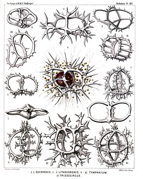
PLATE 82.

Legion NASSELLARIA.

Order STEPHOIDEA.

Families Coronida et Tympanida.


PLATE 82.
Coronida et Tympanida.
Diam. Page.
Fig. 1. Coronidium cervicorne, n. sp., × 400 974
Seen from the apical pole.
Fig. 2. Coronidium acacia, n. sp., × 300 975
Fig. 3. Eucoronis angulata, n. sp., × 400 978
Half from the apical, half from the dorsal side.
Fig. 4. Eucoronis challengeri, n. sp., × 400 978
The red central capsule encloses a large ovate nucleus and is surrounded by numerous xanthellæ.
Fig. 5. Eucoronis nephrospyris, n. sp., × 300 977
Fig. 6. Eucoronis perspicillum, n. sp., × 300 977
Fig. 7. Coronidium dyostephanus, n. sp., × 400 974
Seen from the apical pole.
Fig. 8. Coronidium diadema, n. sp., × 300 974
Fig. 9. Acrocubus octopylus, n. sp., × 300 993
Fig. 10. Parastephanus asymmetricus, n. sp., × 400 1008
Fig. 11. Eutympanium militare, n. sp., × 400 1014
Oblique view.
Fig. 12. Lithocubus astragalus, n. sp., × 400 1012
Fig. 13. Trissocircus globus, n. sp., × 400 986
Plate 82.
Plate 82.


PLATE 83.

Legion NASSELLARIA.

Orders STEPHOIDEA et SPYROIDEA.

Families Stephanida, Semantida, Coronida, Tympanida, Zygospyrida, Phormospyrida et Androspyrida.


PLATE 83.
Stephanida, Semantida, Coronida, Tympanida, Zygospyrida, Phormospyrida et Androspyrida.
Diam. Page.
Fig. 1. Lithotympanum tuberosum, n. sp., × 400 1006
Fig. 2. Eutympanium musicantum, n. sp., × 300 1013
Fig. 3. Semantis distephanus, n. sp., × 300 957
Fig. 4. Sphærospyris globosa, n. sp., × 300 1100
Fig. 5. Trissocyclus stauroporus, n. sp., × 200 987
Fig. 6. Trissocircus binellipsis, n. sp., × 300 985
Fig. 7. Podocoronis toxarium, n. sp., × 200 980
Fig. 8. Androspyris anthropiscus, n. sp., × 400 1093
Fig. 9. Cortina tripus, n. sp., × 200 950
Fig. 10. Cephalospyris cancellata, n. sp., × 400 1035
Fig. 11. Tripospyris furcata, n. sp., × 400 1029
Fig. 12. Petalospyris novena, n. sp., × 400 1062
Basal view of the shell, with the cortinar septum.
Fig. 13. Rhodospyris tricornis, n. sp., × 400 1089
Fig. 14. Desmospyris mammillata, n. sp., × 400 1089
Fig. 15. Phormospyris tricostata, n. sp., × 400 1087
Fig. 16. Zygospyris equus, n. sp., × 300 1056
Fig. 17. Archicircus monostephus, n. sp., × 300 941
Fig. 18. Dipospyris cubus, n. sp., × 400 1036
Basal view of the shell, with the cortinar septum.
Fig. 19. Gamospyris circulus, n. sp., × 200 1042
Fig. 20. Stephanospyris excellens, n. sp., × 300 1043
Plate 83.
Plate 83.


PLATE 84.

Legion NASSELLARIA.

Order SPYROIDEA.

Family Zygospyrida.


PLATE 84.
Zygospyrida.
Diam. Page.
Fig. 1. Tripospyris capitata, n. sp., × 400 1028
Seen from the dorsal side.
Fig. 2. Tripospyris semantis, n. sp., × 300 1026
Seen from the ventral side.
Fig. 3. Tripospyris semantis, n. sp., × 300 1026
Seen from the lateral side.
Fig. 4. Tripospyris eucolpos, n. sp., × 300 1029
Seen from the dorsal side.
Fig. 5. Tripospyris diomma, n. sp., × 400 1026
Half from the right side, half from the basal side.
Fig. 6. Tripospyris cortiniscus, n. sp., × 500 1026
Half from the dorsal, half from the right side.
Fig. 6a. Frontal section through the ring, × 500
Fig. 7. Tripospyris conifera, n. sp., × 400 1027
Seen from the dorsal side.
Fig. 7a. From the basal side, × 200
Fig. 8. Tripospyris euscenium, n. sp. (vel Euscenium tripospyris), × 400 1147
Seen from the frontal or ventral side.
Fig. 9. Triceraspyris gazella, n. sp., × 500 1031
Seen from the ventral side.
Fig. 10. Triceraspyris damæcornis, n. sp., (vel Elaphospyris damæcornis?); compare p. 1032, × 400 1057
Seen from the apical (or basal?) side.
Fig. 11. Triceraspyris giraffa, n. sp., × 400 1031
Seen from the frontal side.
Fig. 12. Triceraspyris corallorrhiza, n. sp., × 400 1031
Seen from the frontal side.
Fig. 13. Tristylospyris scaphipes, n. sp., × 400 1033
Seen from the dorsal side.
Fig. 14. Tristylospyris palmipes, n. sp., × 400 1033
Seen from the dorsal side.
Fig. 15. Tristylospyris clavipes, n. sp., × 400 1033
Seen from the basal side.
Plate 84.
Plate 84.


PLATE 85.

Legion NASSELLARIA.

Order SPYROIDEA.

Family Zygospyrida.


PLATE 85.
Zygospyrida.
Diam. Page.
Fig. 1. Dipospyris forcipata, n. sp., × 300 1037
Fig. 2. Dipospyris irregularis, n. sp., × 200 1037
Fig. 3. Dipospyris chelifer, n. sp., × 300 1037
Fig. 4. Dorcadospyris dinoceras, n. sp., × 400 1041
Fig. 5. Dorcadospyris antilope, n. sp., × 200 1041
Fig. 6. Dorcadospyris dentata, n. sp., × 200 1040
Fig. 7. Dorcadospyris decussata, n. sp., × 200 1041
Fig. 8. Dendrospyris polyrrhiza, n. sp., × 200 1039
Fig. 9. Dendrospyris arborescens, n. sp., × 400 1040
Fig. 10. Stephanospyris cordata, n. sp., × 200 1042
Fig. 11. Stephanospyris verticillata, n. sp., × 300 1043
Plate 85.
Plate 85.


PLATE 86.

Legion NASSELLARIA.

Order SPYROIDEA.

Family Zygospyrida.


PLATE 86.
Zygospyrida.
Diam. Page.
Fig. 1. Ceratospyris polygona, n. sp., × 400 1066
Fig. 2. Ceratospyris strasburgeri, n. sp., × 400 1067
Fig. 3. Ceratospyris allmersii, n. sp., × 400 1067
Fig. 4. Ceratospyris mulderi, n. sp., × 400 1067
Fig. 5. Anthospyris aculeata, n. sp., × 400 1065
Fig. 6. Petalospyris dictyocubus, n. sp., × 400 1063
Fig. 7. Liriospyris hexapoda, n. sp., × 400 1049
Fig. 8. Aegospyris caprina, n. sp., × 400 1054
Fig. 9. Ceratospyris preyeri, n. sp., × 400 1068
Fig. 10. Ceratospyris krausei, n. sp., × 400 1068
Fig. 11. Ceratospyris carnerii, n. sp., × 400 1069
Fig. 12. Elaphospyris alcicornis, n. sp., × 400 1057
Fig. 13. Elaphospyris cervicornis, n. sp., × 400 1057
Plate 86.
Plate 86.


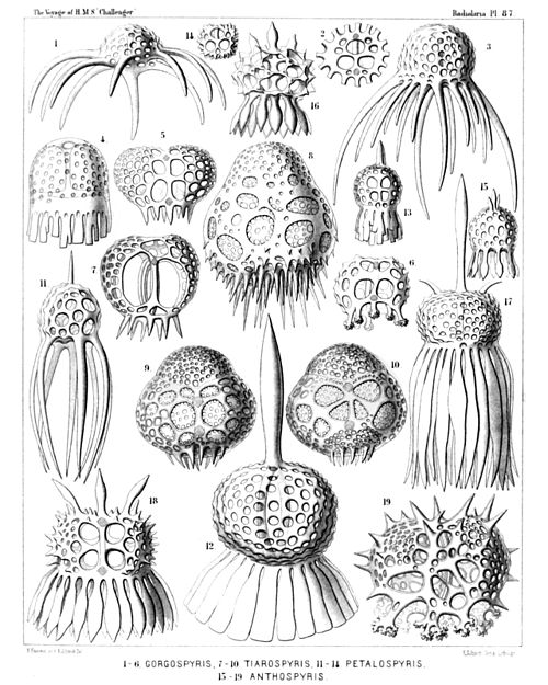
PLATE 87.

Legion NASSELLARIA.

Order SPYROIDEA.

Families Zygospyrida et Tholospyrida.


PLATE 87.
Zygospyrida et Tholospyrida.
Diam. Page.
Fig. 1. Gorgospyris medusa, n. sp., × 300 1070
Fig. 2. Gorgospyris medusetta, n. sp., × 300 1070
From the basal side, with the nine cortinar pores.
Fig. 3. Gorgospyris polypus, n. sp., × 300 1070
Fig. 4. Gorgospyris schizopodia, n. sp., × 400 1071
Fig. 5. Gorgospyris eurycolpos, n. sp., × 300 1071
Fig. 6. Gorgospyris liriope, n. sp., × 300 1071
Fig. 7. Tiarospyris pervia, n. sp., × 400 1082
Fig. 8. Tiarospyris amphora, n. sp., × 400 1083
Fig. 9. Tiarospyris mitra, n. sp., × 400 1082
From the ventral side.
Fig. 10. Tiarospyris mitra, n. sp., × 400 1082
From the dorsal side.
Fig. 11. Petalospyris octopus, n. sp., × 400 1061
Fig. 12. Petalospyris dinoceras, n. sp., × 400 1063
Fig. 13. Petalospyris lobata, n. sp., × 300 1064
Fig. 14. Petalospyris triomma, n. sp., × 200 1060
From the basal side, with the six cortinar pores.
Fig. 15. Anthospyris spathulata, n. sp., × 400 1065
Fig. 16. Anthospyris mammillata, n. sp., × 400 1064
Fig. 17. Anthospyris tragopogon, n. sp., × 300 1066
Fig. 18. Anthospyris doronicum, n. sp., × 300 1065
Fig. 19. Ceratospyris calorrhiza, n. sp., × 400 1069
Plate 87.
Plate 87.


PLATE 88.

Legion NASSELLARIA.

Orders STEPHOIDEA et SPYROIDEA.

Families Tympanida et Androspyrida.


PLATE 88.
Tympanida et Androspyrida.
Diam. Page.
Fig. 1. Toxarium circospyris, n. sp., × 400 995
Fig. 2. Amphispyris sternalis, n. sp., × 300 1096
Fig. 3. Amphispyris costata, n. sp., × 300 1097
Fig. 4. Amphispyris thorax, n. sp., × 300 1096
Fig. 5. Amphispyris subquadrata, n. sp., × 300 1097
Fig. 6. Amphispyris quadrigemina, n. sp., × 300 1096
Fig. 7. Amphispyris toxarium, n. sp., × 300 1097
Fig. 8. Tricolospyris baconiana, n. sp., × 400 1098
Fig. 9. Tricolospyris leibnitziana, n. sp., × 600 1098
Fig. 10. Tricolospyris kantiana, n. sp., × 600 1098
Fig. 11. Tricolospyris newtoniana, n. sp., × 400 1098
Fig. 12. Perispyris lentellipsis, n. sp., × 400 1099
Fig. 13. Perispyris bicincta, n. sp., × 400 1099
Plate 88.
Plate 88.


PLATE 89.

Legion NASSELLARIA.

Order SPYROIDEA.

Families Zygospyrida, Tholospyrida et Androspyrida.


PLATE 89.
Zygospyrida, Tholospyrida et Androspyrida.
Diam. Page.
Fig. 1. Tholospyris tripodiscus, n. sp., × 400 1079
Ventral side.
Fig. 2. Tholospyris fenestrata, n. sp., × 400 1079
Dorsal side.
Fig. 3. Tholospyris ramosa, n. sp., × 400 1079
Dorsal side.
Fig. 4. Tholospyris cupola, n. sp., × 400 1080
Ventral side.
Fig. 5. Therospyris leo, n. sp., × 400 1059
Ventral side.
Fig. 6. Therospyris felis, n. sp., × 400 1059
Dorsal side.
Fig. 7. Dictyospyris stalactites, n. sp., × 400 1073
Ventral side.
Fig. 8. Dictyospyris anthophora, n. sp., × 400 1076
Ventral side.
Fig. 9. Dictyospyris mammillaris, n. sp., × 400 1076
Ventral side.
Fig. 10. Dictyospyris mammillaris, n. sp., × 400 1076
Frontal section.
Fig. 11. Dictyospyris distoma, n. sp., × 300 1073
Ventral side.
Fig. 12. Dictyospyris distoma, n. sp., × 300 1073
Frontal section.
Fig. 13. Lamprospyris darwinii, n. sp., × 300 1094
Ventral side.
Fig. 14. Lamprospyris huxleyi, n. sp., × 300 1094
Ventral side.
Plate 89.
Plate 89.


PLATE 90.

Legion NASSELLARIA.

Order SPYROIDEA.

Family Androspyrida.


PLATE 90.
Androspyrida.
Diam. Page.
Fig. 1. Nephrospyris paradictyum, n. sp. (vel Paradictyum paradoxum), × 250 1102
The complete shell, seen from the frontal side.
Fig. 2. Nephrospyris paradictyum, n. sp., × 250 1102
The incomplete shell, seen from the dorsal side.
Fig. 3. Nephrospyris paradictyum, n. sp., × 500 1102
The sagittal ring, isolated, from the dorsal side; more enlarged.
Fig. 4. Nephrospyris paradictyum, n. sp., × 120 1102
Vertical section through half the shell, exhibiting the thickened margin with the included symbiontes (compare page 1101).
Fig. 5. Nephrospyris paradictyum, n. sp., × 200 1102
Oblique marginal view of the shell.
Fig. 6. Nephrospyris paradictyum, n. sp., × 250 1102
Marginal view of a young specimen, with open fissure between the two parallel net-plates.
Fig. 7. Nephrospyris paradictyum, n. sp., × 250 1102
The soft body alone, without the skeleton. The bilobed central capsule exhibits a central transverse nucleus, and on each lobe a stratum of oil-globules. The kidney-shaped calymma contains on the margin numerous symbiontes (Xanthellæ or Vorticellinæ? Compare page 1102).
Fig. 8. Nephrospyris paradictyum, n. sp., × 500 1102
Three single unicellular symbiontes (Zooxanthellæ?).
Fig. 9. Nephrospyris renilla, n. sp. (vel Nephrodictyum renilla), × 250 1101
The bilobed central capsule is enclosed by the discoidal shell and in the middle constricted by the sagittal ring; it contains a transverse nucleus. The kidney-shaped calymma contains in the peripheral part numerous symbiontes (Xanthellæ or Vorticellinæ? Compare page 1101).
Fig. 10. Nephrospyris renilla, n. sp., × 250 1101
A singular abnormality (occurring not rarely), in which the reduced skeleton has nearly disappeared and the sagittal ring alone remained. The kidney-shaped calymma, however, which encloses numerous symbiontes, has preserved the form of the skeleton. The bilobed central capsule is similar to that in figs. 7 and 9, and is encircled by the thickened sagittal ring.
Plate 90.
Plate 90.