Jump to content

Report on the Radiolaria/Plates11

From Wikisource
1584915Report on the Radiolaria — Plates 101-110Ernst Haeckel

PLATE 101.

Legion PHÆODARIA.

Order PHÆOCYSTINA.

Families Phæodinida, Cannorrhaphida et Aulacanthida.


PLATE 101.
Phæodinida, Cannorrhaphida et Aulacanthida.
Diam. Page.
Fig. 1. Phæocolla primordialis, n. sp., × 300 1544
Central capsule, isolated. The double contoured outer membrane exhibits only one opening, with a radiate operculum and long proboscis. The granular protoplasm encloses clear spherical vacuoles. The sphæroidal nucleus contains irregular amœboid nucleoli.
Fig. 2. Phæodina tripylea, n. sp., × 300 1545
A central capsule in self-division, with two elliptical nuclei. The astropyle is already bisected and has two proboscides.
Fig. 3. Cannorrhaphis spinulosa, n. sp., × 300 1552
A complete specimen with two central capsules, each of which contains two nuclei. The alveolate calymma contains a dark phæodium and is surrounded by tangential tubular needles.
Fig. 4. Cannorrhaphis spinulosa, n. sp., × 300 1552
A single tangential tube.
Fig. 5. Cannorrhaphis spathillata, n. sp., × 300 1552
A single tangential tube.
Fig. 6. Aulactinium actinastrum, n. sp., × 100 1574
A complete specimen, seen in optical meridional section. In the centre the spheroidal central capsule, with its double membrane and three openings (above two lateral parapylæ, below the large astropyle with its radiate operculum). The capsule encloses numerous spherical vacuoles and two hemispherical nuclei, each with numerous nucleoli. The anterior half of the capsule is surrounded by the blackish phæodium. The spherical calymma contains numerous globular alveoles and is pierced by the radial tubes, the proximal ends of which are in contact with the surface of the central capsule (compare Pl. 103, fig. 1).
Fig. 7. Aulactinium actinastrum, n. sp., × 300 1574
A single radial tube.
Fig. 8. Aulactinium actinelium, n. sp., × 200 1574
A single radial tube.
Fig. 9. Mesocena stellata, n. sp., × 600 1557
A single annular piece of the skeleton
Fig. 10. Dictyocha stapedia, n. sp., × 300 1561
A complete specimen, observed living at Ceylon. In the centre is visible the large, spheroidal, tripylean central capsule, with its three openings, containing a large nucleus with numerous nucleoli. Its oral half is covered with the dark phæodium. The voluminous spherical calymma contains numerous globular alveoles and its surface is covered with scattered, stirrup-shaped pieces of the skeleton. Numerous free pseudopodia arise from the surface.
Fig. 11. Dictyocha stapedia, n. sp., × 800 1561
A single piece of the skeleton, from above.
Fig. 12. Dictyocha stapedia, n. sp., × 800 1561
A twin piece of the skeleton.
Fig. 13. Dictyocha medusa, n. sp., × 800 1560
A single piece of the skeleton, from the side.
Fig. 14. Dictyocha medusa, n. sp., × 800 1560
A single piece of the skeleton, from above.
Plate 101.
Plate 101.


PLATE 102.

Legion PHÆODARIA.

Order PHÆOCYSTINA.

Family Aulacanthida.


PLATE 102.
Aulacanthida.
Diam. Page.
Fig. 1. Auloceros elegans, n. sp., × 80 1584
A complete specimen, observed living at Ceylon. In the centre is visible the red central capsule with its three openings, containing a large nucleus of half the size, with numerous nucleoli. The alveolate calymma encloses a green excentric phæodium, is surrounded by a veil of interwoven tangential needles, and forms conical elevations, which enclose the piercing radial tubes. Between these radiate numerous pseudopodia (compare for the single parts, Pl. 103, fig. 1 and Pl. 104, figs. 1-3, and their explanation).
Figs. 2-6. Auloceros furcosus, n. sp., × 100 1583
Distal ends of different radial tubes, exhibiting the great variability of this species.
Fig. 7. Auloceros trigeminus, n. sp., × 300 1584
Distal end of a single tube.
Fig. 8. Auloceros capreolus, n. sp., × 200 1584
Distal End of a Single Tube.
Figs. 9, 10. Auloceros cervinus, n. sp., × 300 1584
Distal ends of two single tubes.
Fig. 12. Auloceros spathillaster, n. sp., × 300 1585
Distal end of a single tube.
Figs. 11, 13. Auloceros arborescens, n. sp., × 300 1585
Distal ends of two single tubes.
Plate 102.
Plate 102.


PLATE 103.

Legion PHÆODARIA.

Order PHÆOCYSTINA.

Family Aulacanthida.


PLATE 103.
Aulacanthida.
Diam. Page.
Fig. 1. Aulographis candelabrum, n. sp., × 100 1583
p, The dark phæodium surrounding the central capsule on its oral part; a, a part of the surrounding alveolate calymma, also surrounding the central capsule; s, the veil of tangential needles covering the surface of the alveolate calymma; r, the big radial tubes, seven of which are visible, with an elegant verticil of terminal branches; f, the numerous pseudopodia radiating between the branches. The central capsule exhibits the following parts:—o, Astropyle; u, parapylæ; e, outer membrane; i, inner membrane; v, vacuoles; n, nucleus; l, nucleoli.
Figs. 2-9. Aulographis pandor, n. sp., × 100 1577
Distal ends of various radial tubes of a single specimen, exhibiting the extraordinary variability of this species.
Fig. 10. Aulographis furcula, n. sp., × 400 1580
A two-branched tube.
Fig. 11. Aulographis furcula, n. sp., × 400 1580
A three-branched tube.
Figs. 12, 13. Aulographis bovicornis, n. sp., × 200 1577
Two tubes with two branches.
Fig. 14. Aulographis bovicornis, n. sp., × 200 1577
A tube with three branches.
Fig. 15. Aulographis triangulum, n. sp., × 200 1580
A single tube.
Fig. 16. Aulographis taumorpha, n. sp., × 300 1577
Two tubes, each with two branches.
Fig. 17. Aulographis triglochin, n. sp., × 300 1578
A tube with three branches.
Figs. 18, 19. Aulographis hexancistra, n. sp., × 300 1581
Distal end of two tubes (one with four, the other with five terminal branches).
Fig. 20. Aulographis dentata, n. sp., × 200 1582
Distal end of a single tube.
Fig. 21. Aulographis ancorata, n. sp., × 300 1578
Two tubes, each with four recurved branches.
Fig. 22. Aulographis tetrancistra, n. sp., × 300 1581
A single tube.
Fig. 23. Aulographis stellata, n. sp., × 300 1578
a and b, Two rudimentary or incompletely developed tubes; c, a well-developed tube of the usual form.
Fig. 24. Aulographis asteriscus, n. sp., × 300 1581
Terminal verticil of a single tube.
Fig. 25. Aulographis cruciata, n. sp., × 300 1578
Distal end of a single tube.
Fig. 26. Aulographis pulvinata, n. sp., × 400 1582
Distal end of a single tube.
Fig. 27. Aulographis serrulata, n. sp., × 400 1582
Distal end of a single tube.
Plate 103.
Plate 103.


PLATE 104.

Legion PHÆODARIA.

Order PHÆOCYSTINA.

Family Aulacanthida.


PLATE 104.
Aulacanthida.
Diam. Page.
Aulacanthida.
Fig. 1. Aulospathis bifurca, n. sp., × 50 1586
A complete specimen, excellently preserved, with an ovate alveolate calymma and two central capsules. The surface of the calymma is covered with tangential needles.
Fig. 2. Aulospathis bifurca, n. sp., × 100 1586
An isolated central capsule of another specimen, surrounded by granules of the phæodium. o, Radiate operculum of the astropyle; u, the two lateral parapylæ; e, external membrane of the capsule; i, internal membrane; c, vacuoles in the protoplasm; n, nucleus; l, numerous nucleoli.
Fig. 3. Aulospathis bifurca, n. sp., × 80 1586
Two central capsules of another specimen, surrounded by the phæodium (Self-division). Characters as in fig. 2.
Fig. 4. Aulospathis bifurca, n. sp., × 100 1586
A single radial tube.
Fig. 5. Aulospathis bifurca, n. sp., × 200 1586
Distal part of another radial tube, partly filled up by air-bubbles.
Fig. 6. Aulospathis trifurca, n. sp., × 200 1586
Distal part of a single radial tube.
Fig. 7. Aulospathis trifurca, n. sp., × 200 1586
Distal part of another radial tube.
Fig. 8. Aulospathis triodon, n. sp., × 100 1587
A single radial tube.
Fig. 9. Aulospathis tetrodon, n. sp., × 200 1588
Distal end of single tube.
Figs. 10-13. Aulospathis polymorpha, n. sp., × 400 1587
Four single terminal branches with very different forms of spathillæ.
Figs. 14-17. Aulospathis variabilis, n. sp., × 400 1588
Four single terminal branches with very different forms of spathillæ.
Plate 104.
Plate 104.


PLATE 105.

Legion PHÆODARIA.

Order PHÆOCYSTINA.

Family Aulacanthida.


PLATE 105.
Aulacanthida.
Diam. Page.
AULACANTHIDA.
Fig. 1. Aulodendron indicum, n. sp., × 200 1590
A single tube.
Fig. 2. Aulodendron pacificum, n. sp., × 400 1589
Distal half of a tube.
Fig. 3. Aulodendron australe, n. sp., × 300 1589
A single tube.
Fig. 4. Aulacantha spinosa, n. sp., × 300 1575
Distal half of a tube.
Fig. 5. Aulodendron antarcticum, n. sp., × 300 1589
A single tube.
Fig. 6. Aulographis pistillum, n. sp., × 300 1579
A single tube.
Fig. 7. Aulographis martagon, n. sp., × 300 1579
Distal end of a single tube.
Fig. 8. Aulographis triæna, n. sp., × 80 1579
A single tube.
Fig. 9. Aulographis flammabunda, n. sp., × 100 1579
Distal end of a tube.
Fig. 10. Aulographis flosculus, n. sp., × 300 1580
Distal end of a tube.
Fig. 11. Aulographis gemmascens, n. sp., × 100 1580
Distal end of a tube.
Fig. 12. Aulographis verticillata, n. sp., × 400 1582
Distal end of a tube.
Fig. 12a. Apical view, with four verticils of five branches.
Fig. 13. Aulographis tripentas, n. sp., × 300 1582
Distal end of a tube.
Fig. 13a. Apical view, with three verticils of five branches.
Fig. 14. Auloceros dicranaster, n. sp., × 400 1585
Distal end of a tube, seen from the side.
Fig. 15. Auloceros dicranaster, n. sp., × 200 1585
Distal end of a tube, seen from the terminal face.
Fig. 16. Aulacantha cannulata, n. sp., × 300 1576
Distal end of a tube.
Plate 105.
Plate 105.


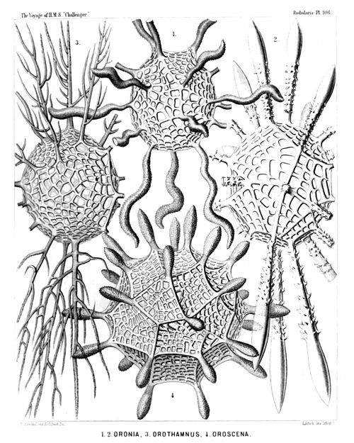
PLATE 106.

Legion PHÆODARIA.

Orders PHÆOSPHÆRIA.

Family Orosphærida.


PLATE 106.
Orosphærida.
Diam. Page.
Orosphærida.
Fig. 1. Orosphæra serpentina, n. sp., × 50 1595
The entire shell.
Fig. 2. Orosphæra horrida, n. sp., × 50 1596
The entire shell.
Fig. 3. Orosphæra arborescens, n. sp. (vel Orothamnus arborescens), × 50 1597
The entire shell.
Fig. 4. Oroscena gegenbauri, n. sp., × 50 1597
The entire shell.
(Compare Pl. 12, fig. 1.)
Plate 106.
Plate 106.


PLATE 107.

Legion PHÆODARIA.

Order PHÆOSPHÆRIA.

Family Orosphærida.


PLATE 107.
Orosphærida.
Diam. Page.
(Fig. 8 of this Plate has no number, by mistake; it is at the top in the middle.)
Fig. 1. Oroplegma diplosphæra, n. sp., × 50 1600
The entire shell, enveloped by an outer mantle of spongy framework.
Fig. 2. Oroplegma giganteum, n. sp., × 200 1601
A small piece of the spongy framework.
Fig. 3. Oroplegma spongiosum, n. sp., × 50 1601
A pyramidal elevation of the inner shell, with its spongy framework, and a radial spine on the top.
Fig. 4. Oroscena bærii, n. sp., × 100 1598
A pyramidal elevation of the shell, with a radial spine on its top.
Fig. 5. Orona maxima, n. sp., × 300 1594
A small piece of the network; the central canals of the bars are partly filled by air.
Fig. 6. Oroscena cuvieri, n. sp., × 50 1598
A single radial spine.
Fig. 7. Orona crassissima, n. sp., × 300 1594
A single bar of the coarse network, with dimpled surface.
Fig. 8. Oroscena mülleri, n. sp., × 50 1598
A single radial spine.
Plate 107.
Plate 107.


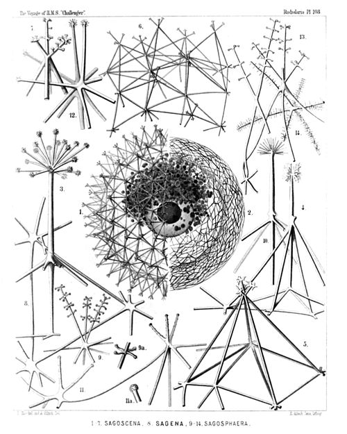
PLATE 108.

Legion PHÆODARIA.

Order PHÆOSPHÆRIA.

Family Sagosphærida.


PLATE 108.
Sagosphærida.
Diam. Page.
Sagosphærida.
Fig. 1. Sagoscena castra, n. sp., × 50 1608
Half the shell, with the enclosed central capsule and the phæodium, stained by carmine. (The central nucleus dark.)
Fig. 2. Sagmarium spongodictyum, n. sp., × 50 1612
Half the shell, with its delicate spongy framework.
Fig. 3. Sagenoscena stellata, n. sp., × 300 1610
Top and axial rod of a pyramid, prolonged into a crowned radial spine.
Fig. 4. Sagenoscena ornata, n. sp., × 300 1610
A single pyramid with its axial rod, prolonged into a crowned radial spine.
Fig. 5. Sagoscena pellorium, n. sp., × 300 1609
A single pyramid of the shell-surface.
Fig. 6. Sagoscena tentorium, n. sp., × 100 1608
A piece of the shell with eight pyramids.
Fig. 7. Sagoscena prætorium, n. sp., × 400 1609
Top of a pyramid.
Fig. 8. Sagena ternaria, n. sp., × 400 1606
A single triangular mesh of the lattice sphere.
Fig. 9. Sagmidium crucicorne, n. sp., × 400 1613
A single nodal point with three radial spines.
Fig. 9a. A portion of a spine, more highly magnified.
Fig. 10. Sagosphæra penicilla, n. sp., × 400 1607
One nodal point and its radial spine.
Fig. 11. Sagosphæra furcilla, n. sp., × 300 1607
Two nodal points of the network.
Fig. 11a. Extremity of a spine.
Fig. 12. Sagmidium quadricorne, n. sp., × 400 1614
A nodal point of the shell surface, with four divergent spines.
Fig. 13. Sagoplegma scenophora, n. sp., × 300 1615
Tops of two pyramids.
Fig. 14. Sagmarium plegmosphærium, n. sp., × 300 1612
A nodal point of the spongy framework.
Plate 108.
Plate 108.


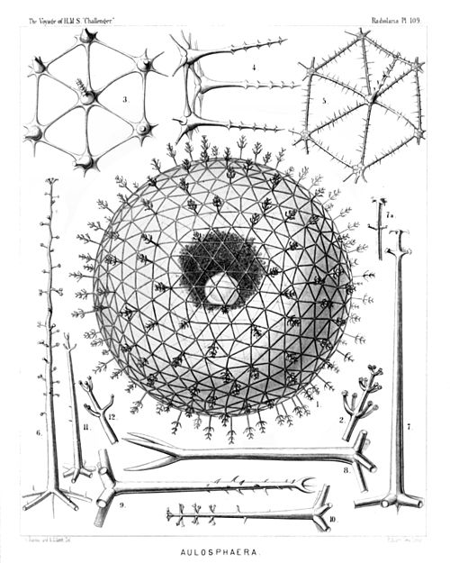
PLATE 109.

Legion PHÆODARIA.

Order PHÆOSPHÆRIA.

Family Aulosphærida.


PLATE 109.
Aulosphærida.
Diam. Page.
Fig. 1. Aulosphæra dendrophora, n. sp., × 50 1625
The entire shell, with the central capsule and its nucleus, enveloped by the dark granular phæodium.
Fig. 2. Aulosphæra dendrophora, n. sp., × 300 1625
A single radial tube.
Fig. 3. Aulosphæra sceptrophora, n. sp., × 300 1625
A hexagonal group of six triangular meshes.
Fig. 4. Aulosphæra sceptrophora, n. sp., × 300 1625
A similar group, seen from the side, with three radial tubes.
Fig. 5. Aulosphæra spinosa, n. sp., × 300 1627
A hexagonal group of six triangular meshes.
Fig. 6. Aulosphæra undulata, n. sp., × 400 1627
A single radial tube.
Fig. 7. Aulosphæra spathillata, n. sp., × 400 1624
A single radial tube.
Fig. 7a. An abnormal variety, × 400
Fig. 8. Aulosphæra triodon, n. sp., × 400 1623
A single radial tube.
Fig. 9. Aulosphæra trifurca, n. sp., × 400 1626
A single radial tube.
Fig. 10. Aulosphæra cruciata, n. sp., × 300 1624
A single radial tube.
Fig. 11. Aulosphæra bisternaria, n. sp., × 300 1624
A single radial tube.
Fig. 12. Aulosphæra bisternaria, n. sp., × 600 1624
Distal end of a single radial tube.
Plate 109.
Plate 109.


PLATE 110.

Legion PHÆODARIA.

Order PHÆOSPHÆRIA.

Family Aulosphærida.


PLATE 110.
Aulosphærida.
Diam. Page.
Fig. 1. Auloscena mirabilis, n. sp., × 50 1628
The complete shell, representing a regular latticed sphere, which is composed of equal hexagonal pyramids; the top of each pyramid bears a radial tube with a terminal corona.
Fig. 2. Auloscena mirabilis, n. sp., × 600 1628
Terminal corona of a single radial tube.
Fig. 3. Auloscena penicillus, n. sp., × 200 1629
A single tent-shaped elevation or six-sided pyramid, bearing on the top a brush-shaped radial tube.
Fig. 4. Auloscena flammabunda, n. sp., × 400 1629
A single radial tube, with a centripetal free prolongation at the base and a verticil of undulate terminal branches at the distal end.
Fig. 5. Auloscena serrata, n. sp., × 600 1630
Terminal corona of a single radial tube.
Fig. 6. Auloscena tentorium, n. sp., × 400 1628
A single radial tube, with a centripetal prolongation at the base and a terminal corona at the distal end.
Fig. 7. Auloscena gigantea, n. sp., × 400 1629
Basal part of a radial tube, exhibiting the internal axial thread and its connection with the six tubes, which form the edges of a flat six-sided pyramid (usually more elevated than the figure exhibits).
Fig. 8. Auloscena spectabilis, n. sp., × 400 1628
Apex of an abnormal pyramid (sometimes occurring), in which seven radial tubes are united, instead of six.
Fig. 9. Auloscena spectabilis, n. sp., × 800 1628
Basal part of a radial tube, in the top of a flat six-sided pyramid; above it the distal part of the same tube with its terminal corona (middle part of the tube wanting).
Fig. 10. Auloscena verticillus, n. sp., × 300 1629
Apex of a six-sided pyramid, seen from the inside.
Fig. 11. Auloscena verticillus, n. sp., × 400 1629
Distal part of a single radial tube, with the terminal corona.
Plate 110.
Plate 110.